Where to start?
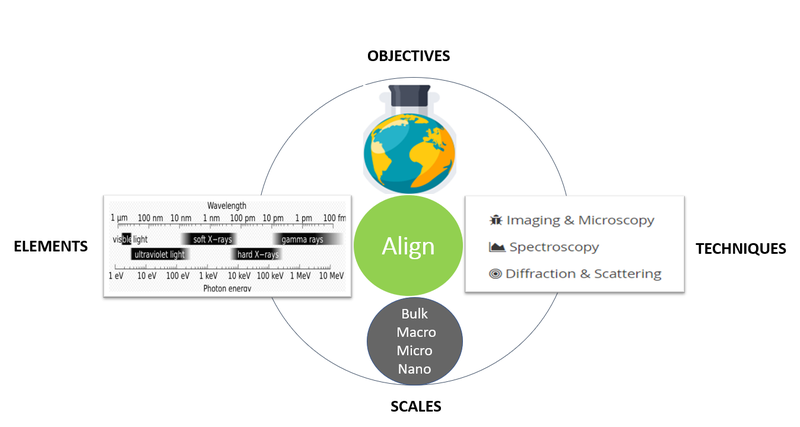
If you are new to synchrotron-based techniques it can be challenging to figure out whether a beamline is suitable for your research. A general approach would be to align the scientific objectives with the main characteristics of a beamline: the absorption energy range of the element(s) of interest, available techniques, and resolution scales.
Understanding the principle of XRI is an important step to decide whether this imaging technique would answer your scientific question. Furthermore, examples of data sets collected at our beamline presented under the Publication/Research Highlights section can be also useful to understand the capabilities of the beamline. In addition, under Readings, there are links to relevant review papers focusing on the principle and application of synchrotron-based X-ray fluorescence imaging in various fields.
In case you are still unsure and you need clarification, please reach out to the beamline staff.
Principle of XRF Imaging
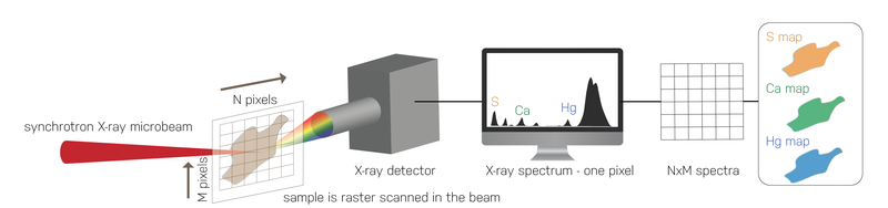
In very simple terms, the beamline components will select, deliver and focus the monochromatic (single wavelength) X-ray light onto the sample mounted in the experimental hutch. The sample is moved either by point-by-point or in continuous fly-scanning mode relative to the incident X-ray light. In the former acquisition mode the sample is moved to a position, stops, then resumes moving; however, in the continuous fly scanning mode data are collected as the sample is moving in both directions. The dwell time for the fly scanning mode can also be set shorter (ms), therefore a collection of data with this mode can be a lot shorter in comparison to the point-by-point scan. The dwell time is how much time the X-ray light is allowed on the specific area or pixel. The size of a pixel is usually consistent with the beam size.
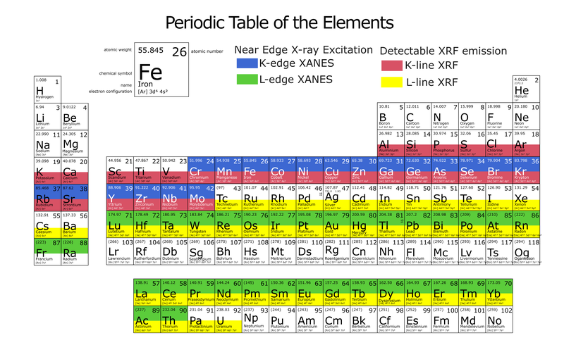
When the incident X-ray photon hits the sample, the inner shell electrons of atoms in the sample become excited, creating photon electrons and a core hole. The vacant inner shell of an atom is rapidly filled with an outer electron, subsequently releasing X-ray fluorescence photon. This emitted X-ray fluorescence photon's energy is unique for each atom, serving as a fingerprint to identify and study the distribution of elements in various types of samples.
The images of elements' distribution in a scanned sample are built by a computer from a single-pixel X-ray fluorescence spectra collected by the X-ray detector. The peaks visible on the computer screen (see image below) would represent the energy of each photon specific to each element detected, the height of the peak indicates number of photons at different energies.
Techniques
X-ray fluorescence imaging
The BioXAS-Imaging beamline operates between 5 and 21 keV energy range. X-ray fluorescence imaging is one of the main techniques with the capability to provide the distribution of elements at a high spatial resolution and at a relatively short acquisition time. The fast bi-directional fly scanning up to 20 ms dwell time, enables scanning large areas and an adequate number of replicates.
In situ micro-XAS
This technique is an excellent complement to the imaging technique as it allows determining speciation at the micro-scale. Once elemental maps are obtained by the XRI technique, selected spots on the map can be targeted with the energy specific to the element of interest. A scan in energy below and above the absorption edge provides information about the oxidation state of the absorbing element. Coupling imaging with micro-XAS is a powerful method to investigate the composition and speciation of various complex matrixes with minimal or no sample preparation requirement.
XAS-imaging
The BioXAS-Imaging beamline has the capability to scan the same map at energy steps of a XANES spectrum.
Available resolution-modes
There are two resolution modes available, the macro-mode with beam size ranging between 20 to 150 um and micro-mode with focused beam size as small as 5 x 5 um. Based on the desired resolution the user can select between the resolution modes. Furthermore, utilization of both resolution modes during a single beamtime is also possible, but this information needs to be stated in the beamline proposal and communicated with the beamline staff.