Users now have access to a new workspace at the beamline where they can inspect/investigate their samples using various microscopes and mount the samples on specifically designed workbench.
The researh team is using XRF-imaging to understand distribution of key elements and biomarkers change in the brain after a stroke. XRF-imaging data of whole body mouse (model organism) section were collected in Macro-mode at 100 micron resolution and 60 ms dwell time.
During the fall shutdown air conditioning system was installed in the BioXAS primary and secondary optics enclosures. The optimum temperature in the enclosures will aid beam stability over time.
During the fall shutdown the BioXAS-Imaging beamline user area received an important upgrade to ensure a quieter workspace area during the beamline operation and data collection.The Imaging beamline received an additional space (setup still in progress), where scientists can inspect/investigate their samples using various microscopes and mount the samples on specifically designed workbench.
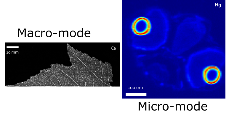
Dont miss the date: Call for proposals opens on January 27, 2021. BioXAS Imaging Macro and Micro resolution modes have recently entered General User phase and accepting proposals. Important information, such as flux, specifications, setup and sample holder sizes for both modes can be found under Resolution modes and User information on this website. Please contact us if you have questions.
Researchers were interested to localize Se contaminant in bumble bees. XRF images were collected with bi-directional fly scanning at 40 μm resolution and 250 ms dwell time. .
Students were interested in how metals/metalloids are distributed within soft tissue of blue mussels (Mytilus edulis).
Our first user at the beamline collected data in micro-mode to investigate phytoremediation potential of plants grown at mining impacted site.
XRF images representing Ca, Zn, Hg and S distribution in zebrafish. Data were collected as fly scan in micro-mode at 5 μm resolution.
Distribution of Ca, Fe and Zn in fern leaves. XRF images collected with bi-directional fly scanning at 100 μm resolution and 50 ms dwell time.
Distribution of Mn and Zn in dry leaves at different resolution. XRF images were collected using the macro-mode setup with bi-directional fly scanning and three different beam size defining apertures (20, 50, 150 microns).
Curiosity driven highschool students investigated Cu mine drainage impact on the surrounding environment. Copper uptake in brown algae (Fucus gardneri) were studied using fly scan XRF imaging in Macro-mode.
Although dry leaves could depict an end, but for us this image meant a beginning, a start to contribute to science and knowledge.