Designed for experiments that require greater/equal resolution to 10 μm. There are two stage-configuration available depending on the sample thickness. The 45 degrees stage-configuration with respect to the incident beam are generally set up for cross-sections varying from a few μms to a couple of hundreds of μms. For thicker samples reaching mm range, the 90 degrees stage-configuration is used.
Specifications
- Beam focused by KB mirrors
- Step-based or bi-directional fly imaging
- Sample stage at either 90 or 45 degress to the incoming beam
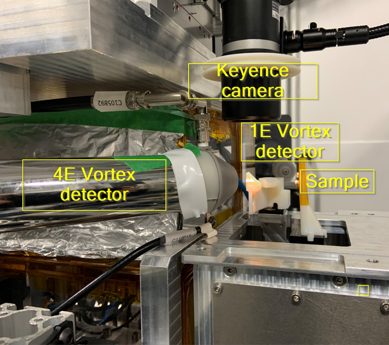
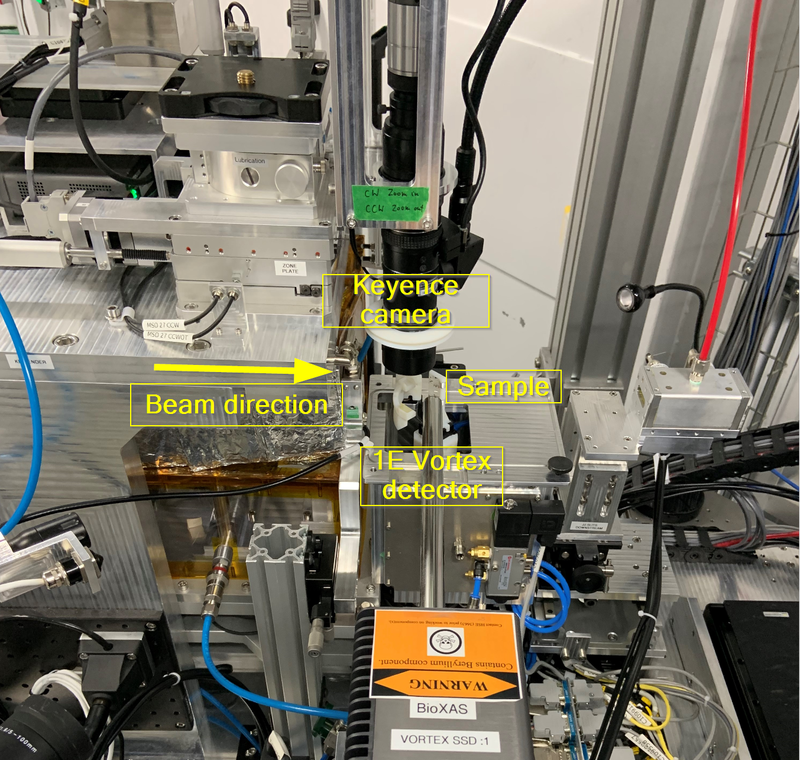
- Keyence camera with long working distance (85 mm), high zoom (variable) lens for sample visualization
- Samples in ambient air or He gas box
- Cryo Jet/LN2 Linkam for sample cooling (to be commissioned)
Beam size and flux
Beam spot sizes measured at 45 degrees (meaning ~1.41X increase in beam H size) and at 10 keV (5th harmonics):
- 10 μm (H) x 5 μm (V) flux 3.2 x 10^11 ph/s/100 mA (full focused beam limited only by the acceptance of the KB mirrors)
- 6 μm (H) x 5 μm (V) flux 1.5 x 10^11ph/s/100 mA (beam cut horizontally at the secondary source)
Setups
Stage configuration set at 90 degrees
- Micro-stage set at 90 degree orientation with and 3E Vortex detector at 45 degrees to the incident beam.
Stage configuration set at 45 degrees
Techniques
- X-ray fluorescence imaging (XFI)
- X-ray absorption spectroscopy (XAS)
- X-ray absorption spectroscopy in situ (u-XAS)
Examples
- Stage configuration at 45 degrees to the incident beam
- The imaging data were collected at a resolution of 5 um at 100 ms dwell time
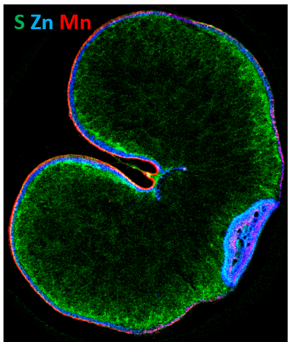
Distribution of Micronutrients in Arborg Oat (Avena sativa L.) Using Synchrotron X-ray Fluorescence Imaging (Deng et al., 2023).